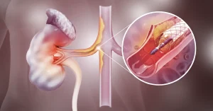
What Are Arteriovenous Malformations?
Arteriovenous malformations (AVMs) are abnormal connections between arteries and veins, bypassing the capillaries — the small blood vessels where oxygen exchange normally occurs.
As a result, blood flows directly from arteries into veins, leading to increased pressure and impaired circulation in the affected area.
Where Can AVMs Occur?
Although AVMs can appear anywhere in the body, the most common and potentially dangerous types are:
- Cerebral AVMs – located in the brain;
- Spinal AVMs – affecting the spinal cord;
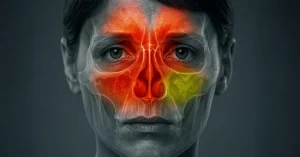
- Peripheral AVMs – found in the limbs, face, chest, or internal organs.
Causes and Risk Factors
Most AVMs are congenital, meaning they are present at birth, even if symptoms appear later in life.
The exact causes are not fully understood, but genetic factors or vascular mutations during fetal development may play a role.
In rare cases, AVMs can be acquired, for example after trauma or surgical procedures.
Symptoms of AVMs
Symptoms vary depending on the location and size of the malformation. Some AVMs cause no symptoms at all.
For cerebral AVMs, common symptoms include:
- Severe or chronic headaches;
- Seizures;
- Weakness or paralysis on one side of the body;
- Vision, speech, or balance problems;
- Confusion or memory loss.
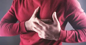
For peripheral AVMs, symptoms may include:
- Swelling, pulsations, or skin discoloration;
- Pain or a burning sensation;
- Ulcers or non-healing wounds;
- Frequent or unexplained bleeding.
Possible Complications
- Hemorrhage (bleeding in the brain or internally) – the most serious complication;
- Stroke (CVA);
- Permanent neurological deficits;
- Heart failure (in extensive AVMs);
- Chronic pain and organ dysfunction.
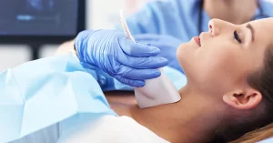
How Are AVMs Diagnosed?
Diagnosis is made using advanced imaging techniques, such as:
- CT (Computed Tomography);
- MRI (Magnetic Resonance Imaging);
- Cerebral or peripheral angiography – the gold standard for visualizing AVMs.
Treatment Options for AVMs
Treatment depends on the type, size, location, and symptoms of the AVM.
Observation and Monitoring
- For small, asymptomatic AVMs, regular monitoring may be recommended.
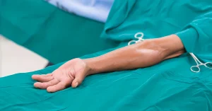
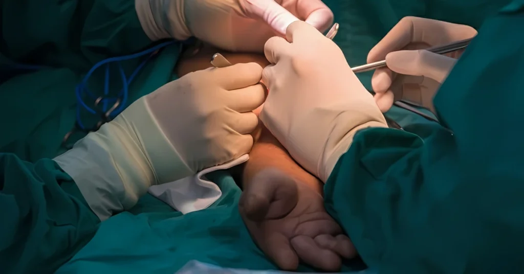
Surgical Treatment
- Surgical resection – complete removal of the AVM when possible;
- Recommended especially for superficial or easily accessible AVMs.
Endovascular Embolization
- A catheter is inserted into the blood vessels to inject a substance that blocks the AVM;
- Often performed before surgery or as a standalone treatment.
Stereotactic Radiosurgery (e.g., Gamma Knife)
- Used for small, deep-seated cerebral AVMs;
- Employs focused radiation to close off the AVM gradually, over 1–3 years.
Living With an Arteriovenous Malformation
An AVM diagnosis does not always mean an emergency, but it requires close medical follow-up.
With appropriate treatment, risks can be significantly reduced. It is important to:
- Follow the monitoring plan set by your neurologist or neurosurgeon;
- Avoid trauma or excessive effort in the affected area;
- Notify your medical team if new or worsening symptoms occur.
When to Seek Emergency Medical Help
Seek immediate medical attention if you experience:
- Sudden, severe headache;
- Speech or vision problems;
- Weakness or numbness on one side of the body;
- Loss of consciousness;
- Any unusual or unexplained bleeding.
Conclusion
Arteriovenous malformations are rare but potentially serious conditions.
With accurate diagnosis and individualized treatment, most patients can lead a normal or near-normal life.
Open communication with your medical specialists and regular monitoring are key to preventing complications.
If you have been diagnosed with an AVM or suspect one, consult a neurologist, neurosurgeon, or interventional radiologist for evaluation and guidance.