The popliteal artery is one of the most important blood vessels in the lower limb. It is located behind the knee and provides blood flow to the calf, muscles, joints, and deep tissues of the leg. Any condition affecting this artery can compromise limb function, mobility, and, in severe cases, the patient’s life.
One of the most common problems encountered at this level is popliteal artery dilation (commonly known as popliteal aneurysm), a pathological widening of the vessel that weakens the arterial wall and can lead to serious complications if not treated in time.
What is the popliteal artery and why is it essential?
The popliteal artery is the continuation of the femoral artery and passes behind the knee through a narrow anatomical space. From here, it divides into two major branches that supply the calves and feet:
- Anterior tibial artery;
- Posterior tibial artery.
The main roles are:
- Transporting oxygenated blood to the calf muscles;
- Maintaining perfusion in the peripheral nerves;
- Vascularization of the knee joint and deep structures;
- Participating in collateral circulation during repetitive knee movements.
Because it is located in an area prone to trauma, mechanical pressure, and repeated bending, the popliteal artery is more vulnerable than other arteries.
What is popliteal vein dilation (popliteal ectasia)?
Popliteal dilation occurs when the arterial wall loses its resistance and the diameter of the vessel increases by more than 50% compared to normal. This enlargement may remain asymptomatic for a long time, but the risks increase with the size of the dilation.
What happens inside the artery:
- The arterial wall becomes thin and fragile;
- Blood flow becomes turbulent;
- Blood clots (thrombi) form;
- These thrombi can migrate to smaller arteries → acute ischemia.
Without treatment, popliteal dilation can lead to:
- Severe ischemia of the limb;
- Tissue necrosis;
- Amputation of the foot;
- Internal bleeding (in case of rupture).

Causes of popliteal artery dilation
The etiology is not entirely clear, but studies show that a combination of factors contributes to the weakening of the arterial wall:
1. Atherosclerosis
This is the most common cause. Fat deposits narrow the artery and increase local pressure, causing pathological widening of the weakened areas.
2. Genetic factors
Some patients have a predisposition to arterial wall fragility.
3. Inflammatory processes
Chronic inflammation degrades the elastic and muscular structures of the arterial wall.
4. Knee trauma
Severe blows, fractures, previous surgery.
5. Connective tissue diseases
For example:
- Marfan syndrome;
- Ehlers-Danlos syndrome;
- Vasculitis.
6. Lifestyle/cardiovascular risk factors
- Smoking;
- High blood pressure;
- High cholesterol;
- Diabetes;
- Age over 60;
- Male gender.
Symptoms of popliteal artery dilation
Many popliteal dilations are asymptomatic and are discovered accidentally during ultrasound or MRI scans. However, when symptoms do occur, they should not be ignored:
Common signs:
- Pulse or pressure sensation in the popliteal artery;
- Dull pain behind the knee;
- Swelling of the calf due to compression of the veins;
- Numbness or tingling;
- Decreased muscle strength;
- Cold feet or paleness during exercise.
Warning signs (medical emergency):
- Intense and sudden pain in the calf;
- Cold, blue, or numb foot;
- Inability to move the toes;
- Disappearance of the pulse at the ankle;
- Ulcers or wounds that do not heal.
How is popliteal dilation diagnosed?
The diagnosis is based on clinical examination and imaging investigations:
1. Doppler ultrasound
- Measures artery diameter;
- Detects blood clots;
- Assesses blood flow.
2. CT angiography / MRI angiography
Useful when planning surgery.
3. Clinical examination
- Palpation of the popliteal pulse;
- Assessment of foot temperature and coloration;
- Identification of a pulsatile mass.
Early diagnosis reduces the risk of amputation by more than 70%.
When should you see a doctor?
- You notice a lump behind your knee;
- You have persistent pain in your calf;
- You have symptoms of ischemia;
- You have a family history of vascular problems;
- You are a smoker and have pain when walking.

It is essential that patients with unilateral popliteal dilation also be examined at the abdominal level, as there is an increased risk of associated aortic aneurysm.
Treatment of popliteal artery dilation
Treatment depends on size, symptoms, and risk of complications.
Monitoring (for small, asymptomatic dilations)
- Ultrasound scan at 6–12 months;
- Blood pressure monitoring;
- Smoking cessation;
- Statins/antiplatelet agents.
Arterial bypass (traditional treatment)
The internal saphenous vein is harvested and used to create a bypass that circumvents the affected segment. This method has the best long-term results.
Ligation of the dilation + bypass
The dilated segment is excluded from circulation.
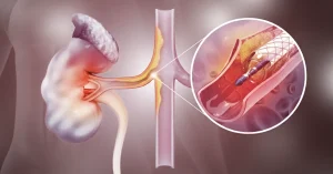
Endovascular treatment (minimally invasive)
- Implantation of a stent graft;
- Lower risk in elderly patients;
- Rapid recovery.
It is ideal in complicated dilations or in patients with increased surgical risk.
Emergency treatment
If the dilation fills with clots and blocks the flow:
- Thrombolysis;
- Immediate bypass;
- Limb salvage within the first 6 hours.
Early intervention is essential to maintain leg function.
Frequently Asked Questions
They are the same condition: the correct medical term is “popliteal aneurysm,” while “popliteal dilation” is the more accessible term used in practice. It occurs when the artery’s diameter increases by more than 50% compared to normal.
No. Without treatment, the arterial wall continues to weaken, and the risk of thrombosis, ischemia, or amputation increases as the dilation grows.
Endovascular intervention for the popliteal artery starts from €5,500. The exact cost depends on the complexity of each individual case. Give us a call and we’ll figure out the next steps together. Schedule a Consultation.
The decision depends on the size of the dilation and your symptoms. Small, asymptomatic dilations are monitored with ultrasound every 6-12 months. Symptomatic ones or those exceeding a certain diameter require surgical or endovascular intervention.
Arterial bypass is the classic procedure, involving an incision and a longer recovery, but with excellent long-term results. A stent-graft is minimally invasive, with rapid recovery, and is especially recommended for older patients or those with higher surgical risk.
It depends on the type of intervention. After a classic bypass, full recovery takes several weeks. After endovascular treatment, patients are typically discharged within 1-2 days and resume normal activities much sooner.
Yes. Patients with a popliteal aneurysm have an increased risk of an associated abdominal aortic aneurysm. Your vascular surgeon will recommend additional investigations to rule this out.
A vascular surgeon. They will perform a clinical evaluation, request a Doppler ultrasound, and if needed, an Angio-CT scan to establish the diagnosis and treatment plan.
Conclusion
The popliteal artery plays an essential role in supplying blood to the lower limbs. Popliteal artery dilation is a serious condition with a high risk of thrombosis, ischemia, and amputation, but it is completely treatable if diagnosed early. Modern interventions, whether surgical or endovascular, offer excellent results when the patient sees a doctor in the early stages.
If there is pain, pulsating mass, or any symptom behind the knee, vascular evaluation should not be delayed.